[ISSUE CoM]AI-assisted lung cancer digital pathology image analysis – TMU’s research paper honorably published in well-known international journal
After receiving his Ph.D. degree in the Institute of Basic Medical Sciences of National Cheng Kung University in July, 2013, Dr. Lee, Yu-Cheng started his post-doctoral research in Baylor College of Medicine in Texas, USA, focusing on the relation between Extracellular Matrix (ECM) and metastatic bladder cancer as well as the construction of patient-derived xenograft (PDX) animal models. Lee started teaching in Taipei Medical University in September, 2019, and currently serves as an assistant professor in the Graduate Institute of Medical Science, College of Medicine.
Construction and Application of Large-scale Image Databases, a project conducted by Taipei Medical University (TMU) and subsidized by the Ministry of Science and Technology (MOST), has brought significant benefits to the clinical practice. The research paper on this project got published in 2021 in Nature Communications, which is indeed a remarkable achievement. It marks the first key accomplishment of TMU President Chien-Huang Lin, for his relentless efforts over the past 3 years in facilitating the development of digital pathology and initiating the digitization of pathology slides in Taiwan’s top 10 leading causes of cancer death.
Leveraging the supercomputer “TAIWANIA 2” deployed by National Center for High-Performance Computing, NARLabs under the Forward-looking Infrastructure Plan, the AI system co-developed by TMU and aetherAI is a world-leading whole-slide pathology image recognition system for lung cancer that does not require manual annotation. The system can distinguish between benign and malignant tumors with an accuracy rate of over 95%. More importantly, the time required for recognition is greatly reduced by 2/3, accelerating the pathological diagnosis procedure and allowing more time for treatment.
As informed by TMU Vice President Cheng-Yu Chen, the research paper titled “An annotation-free whole-slide training approach to pathological classification of lung cancer types using deep learning” was published in Nature Communications, a well-known international medical journal, on February 19, 2021 (https://www.nature.com/articles/s41467-021-21467-y). The research team collected pathology slides of lung tumor tissues over the past 20 years from Taipei Medical University Hospital, Taipei Municipal Wanfang Hospital, and Shuang Ho Hospital. The slides were scanned and digitized to build a massive database, further contributing to the digitization of pathology.
Professor Chi-Long Chen from the Department of Pathology, School of Medicine, TMU and Chi-Chung Chen, an engineer at aetherAI are the lead authors on the paper, while Cheng-Yu Chen, TMU Vice President and Chao-Yuan Yeh, CEO at aetherAI are the corresponding authors. As advised by Professor Chi-Long Chen, former Vice Dean of College of Medicine, these pathology slides used in the research were made from lung tumor tissues extracted through biopsy or surgery after X-ray, NMR or computed tomography examinations. A total of over 9,000 slides were scanned into digital image files for pathologists to annotate the cancerous and non-cancerous areas. The annotated results were used to train the AI model. After constant training and corrections, the system has achieved a high accuracy rate at around 95%.
The conventional diagnosis method for lung cancer is conducted by delivering the potential tumor tissues extracted through biopsy or surgery to the Pathology and Laboratory Department, where they are made into pathology slides for pathologists to examine through a microscope. Not only is this method time-consuming and laborious, the results may also vary depending on the experience level of the pathologist conducting the examination. Moreover, during manual analysis, pathologists need to first annotate potential tumor areas, then further perform substantial annotation and diagnoses before they can get a conclusion. By contrast, the whole-slide-based AI system developed by TMU and aetherAI can identify lung cancer and tumor sub-types without any manual annotation involved.
According to Professor Chi-Long Chen, some other countries have developed similar recognition systems, but they require splitting a pathology slide into tens of thousands of image files and having them annotated by pathologists before used for AI training. Not only is such method subject to the availability of professional pathologists, its accuracy may also be compromised due to the overlapping of images, thus affecting the final analysis.
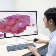
In contrast, under the world-leading whole-slide pathology image recognition system for lung cancer co-developed by TMU and aetherAI, AI learns from the digital images of pathology slides the way pathologists examine the slides through a microscope. This approach has reduced image overlaps and improved the accuracy of the analysis. According to Professor Chen, the system first distinguishes normal from abnormal areas in the digital pathology slide images of lung cancer, then it analyzes whether the tumor tissues in the abnormal areas are benign or malignant. If they are malignant, they can be further classified into lung adenocarcinoma or squamous cell carcinoma of the lung. Finally, the AI analysis results are delivered to pathologists for final diagnostic confirmation. 【Picture : AI assists with pathological diagnoses – the red patches indicate AI-predicted tumor areas】

As told by Professor Chen, the AI analysis of pathology slides serve as a preliminary diagnosis for pathologists, as it not only indicates the potential tumor areas, but also analyzes whether the tumor is benign or malignant. With that done, pathologists only need to conduct further examination on the areas flagged by the system before making final diagnoses. The system has reduced the possibility of human misjudgment, and even accelerated the whole diagnostic process. For example, there are generally 8 to 15 pathology slides from a patient. If the conventional method is adopted, it will take approximately 10 to 15 minutes for pathologists to examine these slides, but with the assistance of AI analysis, the time required is shortened by 2/3 to approximately 3 to 5 minutes.
According to the latest statistics from the Ministry of Health and Welfare, the cancer death rate in Taiwan reached 212.9 per 100,000 persons in 2019, and lung cancer is the deadliest cancer among the top 10 leading causes of cancer death, following by liver cancer, colorectal cancer, anal cancer, female breast cancer, oral cancer, male prostate cancer, pancreas cancer, stomach cancer, esophageal cancer, and ovarian cancer. By the end of 2020, lung cancer has become the most common cause of cancer death in the world, and unquestionably the biggest threat to people’s health. Given the rising incidence rates of lung cancer in Taiwan over the years and the fact that early-stage symptoms can be easily neglected, Professor Chen believes that early diagnosis and treatment can improve the situation and save lives.
 Prof. Chi-Long Chen
International Master/Ph.D. Program in Medicine
Research Interests:
Pathology
Oncologic Pathology
Novel biomarkers in cancers
Email: chencl@tmu.edu.tw


 Total Users : 354317
